We coducted a motility test. Positive motility= bacteria spreads from stab
-used asceptic technique to dip a sterile inoculating needle into a broth culture.
-stab the bacteria into the center of the test medium to about halfway or three quarters depth
-incubate at 30'C for 24-48 hours
Bacterial Smear:
1-place a loopful of distilled water in the center of the slide
2-transfer a small amount of culture from the agar surface into the water drop using a sterile loop. Spread the mixture into a thin film
3-Transfer a loopful of liquid culture to the center of the slide using a sterile loop
4-spread the drop of culture into a thin film
5-flame the loop before putting it down
6-allow the smear to air dry
7-pass the slide quickly through flame three times, smear slide up
8-place the slide on a slide warmer(60'C for 10minutes)
9-add 95% methanol to the dried smear for at least 1 minute. Rinse off the methanol
10-Roll the cotton swab in the center of the slide
11-allow the smear to air dry
12-fix the smear by heating
Simple Stain:
1-Cover the fixed smear with several drops of stain
2-rinse the slide with water to remove excess stain
3-blot water from the slide with bibulous paper
4-examine the stained smear under the microscope using oil immersion lens
Gram Stain:
-Place a fixed smear on a rack over a staining tray or sink
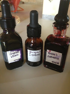
-Cover the smear with crystal violet for 20 Seconds
-Rinse the slide with water to remove excess crystal violet
-Cover the smear with Gram's iodine for 1 minute
-Rinse the slide with water to remove excess iodine solution
-decolorize with 95% ethanol or ethanol/acetone. Hold slide at 45' angle while adding decolorizing reagent drop by drop until color stops running
-imediately rinse slide to remove decolorizing agent
-cover smear with safranin for 1 minute
-rinse slide with water to remove excess safranin
-blot water from slide with pieces of bibulous paper
After Examining the smear under the microscope using the oil immersion lens, we concluded our bacteria is Gram Positive
Tuesday, May 28, 2013
May 16 Day 3
Show growth of broth, slant, agar plate(colonies)
There was great growth at the top of the agar slant:
There was growth in colonies of the agar plate:
We flicked our broth tube, and bacteria in large and small chunks floated, showing there was growth:
WE placed the broth, slant, and agar plates in the refrigerator so the bacteria will not keep growing, or die. Refrigeration just pauses the growth of the bacteria.
We looked at our bacteria through the microscope and saw that it grows in colonies:
at 40x power, using oil(oil emersion lens) for a maximum, "up close" view.
We concluded our bacteria is bacillus, or rod shaped:
Dr. P did an experiment to show that bacteriais killed by bacteriophage. Dr. P wrote Letters with bacteriophage. You can see it killed the bacteria, because no bacteria is present on the letters.
The results are:
May 28, 29, and 30 - Day 10, Day 11, and Day 12
Selective and Differenctial Media
Purpose: Isolate bacteria based on their salt tolerance and differentiate among theses isolates for mannitol fermentation.
Materials: Mannitol salt agar plate, blood plate, E and B plate, Phenol Alcohol plate, MacConkey plate and, Unknown bacteria in a slant

Procedure: For each plate:
1. Sterilize the loop and then gathered some bacteria from our original bacteria from the slant.
2. For each slant, Maggie inoculated each plate with a smear from our original bacteria.
3. Then we put each plate into the 30 degrees C incubator.
4. It will remain in the incubator for 24 hours.
Results:
Blood Plate- Some Growth, Gamma Hemolysis

Mannitol Salt Agar Plate- No Growth

Phenol Alcohol Plate- Growth

MacConkey Plate- No Growth

E and B Plate-No Growth

MRSA Test and Strep Throat Bacteria Test
Materials needed: Saline Solution, Swabs, a throat to swab, a nose to swab, Blood Agar Plate,
Procedure: (Strep Test)

1. Get a Blood Agar Plate
2. Swab a throat
3. Spread the bacteria on the plate
4. Put bacitracin on the plate
5. Put the Plate into 37 degrees C incubator
6. Look for lysis of the blood

Results: Much bacteria grew, Alpha Hemolysis
Procedure: (MRSA Test, Nose)

1. Get a Mannitol Salt Agar Plate
2. Dip a swab into the saline solution
3. Swab both sides of the nose
4. Spread what was gathered from the nose onto the Mannitol Salt Agar Plate
5. Spread bacitracin on the plate
6.Place plate into the 37 degree incubator
Results: Bacteria grew yellow in color, therefore positive for S. aureus.
Antibiotic Testing
Materials needed: Antibiotic disks: (Penicillin, Erythromycin, Tetracycline, Neomycin, and Chloramphenicol), Nutrient Agar Plate, Swab, Unknown Bacteria, Flame, Forceps
Procedure:
1. Get a Nutrient Agar Plate
2. Put Forceps into alcohol for purification
3. Differentiate 5 spots in the Agar plate for 5 different antibiotics: Penicillin, Erythromycin, Tetracycline, Neomycin, and Chloramphenicol.
4. Get a swab and gather unknown bacteria from the slant of bacteria and spread the bacteria all over the Nutrient Agar Plate.
5. Put the forceps into the alcohol then into the flame in order to sterilize them.
6. Pick up an antibiotic disk and place on the specified area in the Nutrient Agar Plate.
7. Put Plate into the 30 degrees C incubator
** Repeat Steps 5 and 6 until all antibiotic disks are placed on the Agar Plate.

Results: Penicillin, Erythromycin, Neomycin, and Chloramphenicol all killed the bacteria. Tetracycline, did kill some of the bacteria, but not entirely or as well as the other four did.
Result of Our Unknown Bacteria:

B. subtilis
-Gram Positive
-Bacilli
-Motile
-Aerobic
-Gaseous
-Endospores
Immunodetective BioKit: Antibody- Antigen Reaction in Agar
Procedure:
1. Get a petri dish divide it into 3 sections
2. In one of the sections make 3 wells and fill them with various solutions:
Well 1: Goat Anti-swine Albumin
Well 2: Goat Anti-horse Albumin
Well 3: Goat Anti-bowvine Albumin
Well 4: Swine Albumin
2. Replace and and cover the dish at room temperature.
3. Record results after 16-48 hrs
1. Get an Elisa plate and label each section
 2. Add antigen (green tube) to wells of microplate strip.
3. Incubate for 5 minutes
4.Wash off the strip with wash buffer
5. Add the serum (yellow tube), positive (purple tube), and negative control (clear tube).
6. Wait 5 minutes
7. Wash it again
8. Use enzyme (orange tube) to detect antigen- add it to each section
9. Wait 5 minutes
10.Wash it again and again
11.Add SUB (brown tube) into each section
12. Wait 5 minutes
13. Observe to see if there is a color change- from colorless to blue. If there is a color change it is positive
Results:
The positive control turned blue meaning it has antibodies towards this antigen (AIDS). The other two controls did not change in color meaning they do not have antibodies towards the antigen.

UV Light
-There is a mixture of all the bacteria in our class.
-Use UV rays to treat it: a UV ray wand was used by stirring and sitting in the beaker of bacteria.
Yogurt
-Boil milk till foaming
-Cool milk to room temperature
-Add spoonful of yogurt to the milk
-Incubate it in37 degrees C
Selective and Differenctial Media
Purpose: Isolate bacteria based on their salt tolerance and differentiate among theses isolates for mannitol fermentation.
Materials: Mannitol salt agar plate, blood plate, E and B plate, Phenol Alcohol plate, MacConkey plate and, Unknown bacteria in a slant

Procedure: For each plate:
1. Sterilize the loop and then gathered some bacteria from our original bacteria from the slant.
2. For each slant, Maggie inoculated each plate with a smear from our original bacteria.
3. Then we put each plate into the 30 degrees C incubator.
4. It will remain in the incubator for 24 hours.
Results:
Blood Plate- Some Growth, Gamma Hemolysis

Mannitol Salt Agar Plate- No Growth

Phenol Alcohol Plate- Growth

MacConkey Plate- No Growth

E and B Plate-No Growth

MRSA Test and Strep Throat Bacteria Test
Materials needed: Saline Solution, Swabs, a throat to swab, a nose to swab, Blood Agar Plate,
Procedure: (Strep Test)

1. Get a Blood Agar Plate
2. Swab a throat
3. Spread the bacteria on the plate
4. Put bacitracin on the plate
5. Put the Plate into 37 degrees C incubator
6. Look for lysis of the blood

Results: Much bacteria grew, Alpha Hemolysis
Procedure: (MRSA Test, Nose)

1. Get a Mannitol Salt Agar Plate
2. Dip a swab into the saline solution
3. Swab both sides of the nose
4. Spread what was gathered from the nose onto the Mannitol Salt Agar Plate
5. Spread bacitracin on the plate
6.Place plate into the 37 degree incubator
Results: Bacteria grew yellow in color, therefore positive for S. aureus.
Antibiotic Testing
Materials needed: Antibiotic disks: (Penicillin, Erythromycin, Tetracycline, Neomycin, and Chloramphenicol), Nutrient Agar Plate, Swab, Unknown Bacteria, Flame, Forceps
Procedure:
1. Get a Nutrient Agar Plate
2. Put Forceps into alcohol for purification
3. Differentiate 5 spots in the Agar plate for 5 different antibiotics: Penicillin, Erythromycin, Tetracycline, Neomycin, and Chloramphenicol.
4. Get a swab and gather unknown bacteria from the slant of bacteria and spread the bacteria all over the Nutrient Agar Plate.
5. Put the forceps into the alcohol then into the flame in order to sterilize them.
6. Pick up an antibiotic disk and place on the specified area in the Nutrient Agar Plate.
7. Put Plate into the 30 degrees C incubator
** Repeat Steps 5 and 6 until all antibiotic disks are placed on the Agar Plate.

Results: Penicillin, Erythromycin, Neomycin, and Chloramphenicol all killed the bacteria. Tetracycline, did kill some of the bacteria, but not entirely or as well as the other four did.
Result of Our Unknown Bacteria:

B. subtilis
-Gram Positive
-Bacilli
-Motile
-Aerobic
-Gaseous
-Endospores
Immunodetective BioKit: Antibody- Antigen Reaction in Agar
Procedure:
1. Get a petri dish divide it into 3 sections
2. In one of the sections make 3 wells and fill them with various solutions:
Well 1: Goat Anti-swine Albumin
Well 2: Goat Anti-horse Albumin
Well 3: Goat Anti-bowvine Albumin
Well 4: Swine Albumin
2. Replace and and cover the dish at room temperature.
3. Record results after 16-48 hrs
1. Get an Elisa plate and label each section
 2. Add antigen (green tube) to wells of microplate strip.
3. Incubate for 5 minutes
4.Wash off the strip with wash buffer
5. Add the serum (yellow tube), positive (purple tube), and negative control (clear tube).
6. Wait 5 minutes
7. Wash it again
8. Use enzyme (orange tube) to detect antigen- add it to each section
9. Wait 5 minutes
10.Wash it again and again
11.Add SUB (brown tube) into each section
12. Wait 5 minutes
13. Observe to see if there is a color change- from colorless to blue. If there is a color change it is positive
Results:
The positive control turned blue meaning it has antibodies towards this antigen (AIDS). The other two controls did not change in color meaning they do not have antibodies towards the antigen.

UV Light
-There is a mixture of all the bacteria in our class.
-Use UV rays to treat it: a UV ray wand was used by stirring and sitting in the beaker of bacteria.
Yogurt
-Boil milk till foaming
-Cool milk to room temperature
-Add spoonful of yogurt to the milk
-Incubate it in37 degrees C
Wednesday, May 22, 2013
May 15 Day 2
Environmental samples results
Fountain:
Maura's Tongue:
Today we received our unknown bacteria!
Dr. Pathakamuri told us that inoculated tubes and plates of this bacteria grow in the 30'C incubator.
We prepared bacteria cultures in a broth tube, an Agar slant, and an agar plate. We first used the asceptic technique for inoculating each.
Broth tube:
We used the asceptic technique for sterilization, then took a loop to the unknown and and inserted it into the fresh broth to grow overnight.

Lastly, Dr. P added bacteriophages to a. agar plate with bacteria that has already grown in colonies. He incubated it. We will check the results tomorrow to see if the bacteriophages do kill bacteria.
Monday, May 20, 2013
May 14, First Day of Lab
Tuesday May 14
First Day of Lab
Lab Safety Rules, Asceptic Technique, Microscope, Sterilization, preparing culture media, Sample Collection, Spread Plate
We learned Microscope Magnification. We will mostly be using 400X magnification
http://www.cas.miamioh.edu/mbi-ws/microscopes/Magnification.html
Learned Importance of Safety
-to reduce the risk of infection and prevent potentially harmful microbes from leaving the laboratory
Proper safety procedure for the start of each day in the lab includes putting on a lab coat, washing hands throughly with antibacterial soap, and decontaminating the workbench with disinfectant.
Joseph Lister first introduced carbolic acid, now called phenol, to sterilise surgical instruments. We still use a form of phenol (2% phenol) as the lab disinfectant today.
Before washing our hands, Dr. Pathakamuri had us put our thumbprint on an agar plate
Agar helps bacteria grow.
After washing our hands, we put our thumbprint on a different section of the agar plate. We put the plate in the 25'C incubator.
We took two sample bacteria's: the first from the back of Maura Bobak's tongue, and the second from the water fountain on first floor of Cosmas and Damian Building at Franciscan University. We used a cotton swab for both.
After obtaining a sample from the fountain piece, we learned how to properly spread the bacteria on an agar plate with the cotton swab, shown below.
This spreading technique will be used in future tests.
Our thumbprint and Environmental Agar plates were both labeled and placed in the 25'C incubator.
Attention: Samples from our microbiology lab are to be put on the nursing majors shelf, not the biology majors shelf, or we're in big trouble.
As we left the lab, We cleared our lab bench, sprayed and wiped down the lab bench with 2% phenol, washed our hands thoroughly, and hung up our lab coats. Nothing was left in the lab, or taken from the lab out. This is standard procedure for each day.
First Day of Lab
Lab Safety Rules, Asceptic Technique, Microscope, Sterilization, preparing culture media, Sample Collection, Spread Plate
We learned Microscope Magnification. We will mostly be using 400X magnification
http://www.cas.miamioh.edu/mbi-ws/microscopes/Magnification.html
Learned Importance of Safety
-to reduce the risk of infection and prevent potentially harmful microbes from leaving the laboratory
Proper safety procedure for the start of each day in the lab includes putting on a lab coat, washing hands throughly with antibacterial soap, and decontaminating the workbench with disinfectant.
Joseph Lister first introduced carbolic acid, now called phenol, to sterilise surgical instruments. We still use a form of phenol (2% phenol) as the lab disinfectant today.
Before washing our hands, Dr. Pathakamuri had us put our thumbprint on an agar plate
Agar helps bacteria grow.
After washing our hands, we put our thumbprint on a different section of the agar plate. We put the plate in the 25'C incubator.
Next, we learned the Aseptic Technique.
This technique came from Joseph Lister.
We first lit the bunsen burner, put our hook tool above the dark blue flame to turn bright red all the way through to thoroughly decontaminate the hook. Next, we opened a tube, held it above the dark blue flame right side up, on a slight slant to decontaminate the top. We then practiced putting the hook tool inside, decontaminating and covering the top of the tube, and decontaminating the hook once again.
Next, we took bacteria samples using cotton swabs from a place in the environment of our choosing to grow on an agar plate.
We took two sample bacteria's: the first from the back of Maura Bobak's tongue, and the second from the water fountain on first floor of Cosmas and Damian Building at Franciscan University. We used a cotton swab for both.
After obtaining a sample from the fountain piece, we learned how to properly spread the bacteria on an agar plate with the cotton swab, shown below.
This spreading technique will be used in future tests.
Our thumbprint and Environmental Agar plates were both labeled and placed in the 25'C incubator.
Attention: Samples from our microbiology lab are to be put on the nursing majors shelf, not the biology majors shelf, or we're in big trouble.
As we left the lab, We cleared our lab bench, sprayed and wiped down the lab bench with 2% phenol, washed our hands thoroughly, and hung up our lab coats. Nothing was left in the lab, or taken from the lab out. This is standard procedure for each day.
Subscribe to:
Posts (Atom)


