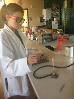
Monday, May 20, Day 5
Smearing and Staining
Preparing a Bacterial Smear
1. Write name of bacterium on one end of the slide.
2.
Place a loopful of distilled water in the center
of the slide.
3.
Transfer a small amount of culture from the agar
surface into the water drop using a sterile loop. Spread the mixture into a
thin film. And allow the smear to air dry.
4.
Pass the slide through the flame three times,
with the smear side up. The smear is now “heat fixed.”
5.
Add 95% methanol to the dried smear for about
one minute and then rinse off with distilled water.
Preparing a Simple Stain
1.
Cover fixed smear with several drops of stain
for the times indicated below:
Stain
|
Time
|
Carbolfuchsin
|
5-10 seconds
|
Crystal violet
|
20-30 seconds
|
Methylene blue
|
At least 1 minute
|
Safranin
|
At least 1 minute
|
2.
Rinse the slide with water to remove excess
stain, blot with bibulous paper.
3.
Examine under microscope using the oil immersion
lens.
Preparing a Gram Stain
1.
Place slide with a fixed smear on rack over
sink.
2.
Cover smear with crystal violet for 20 seconds.
3.
Rinse the slide with water to remove excess
crystal violet.
4.
Cover the smear with Gram’s iodine for one
minute.
5.
Rinse the slide with water to remove excess iodine
solution.
6.
Decolorize with 95% ethanol/acetone. Hold slide
at a 45-degree angle while adding decolorizing reagent drop by drop until color
stops running.
7.
Immediately rinse the slide to remove the
decolorizing agent.
8.
Cover the smear with safranin for one minute.
9.
Rinse the slide with water to remove excess
safranin.
10. Blot
with bibulous paper.
11. Examine
under microscope using the oil immersion lens.
Preparing a Negative Stain
1.
Place a small drop if nigrosin near one end of a
clean slide.
2.
Use a sterilized inoculating loop to transfer a
small amount of bacterial from an agar surface or a loopful of broth culture
into the nigrosin drop. Mix well within that small diameter.
3.
Touch the short edge of another clean microscope
slide at a 30-degree angle in the bacteria-nigrosin drop.
4.
The resulting smear should include a thin film
with a feathered edge. Allow to dry completely.
5.
Examine under microscope using the oil immersion
lens.
Tuesday, May 21, Day 5
Staining
Preparing an Endospore Stain
1.
Place the slide on a beaker with boiling water.
2.
Place paper on the slide and saturate the paper
with malachite green. Stain for 5-6 minutes. Add additional stain as it
evaporates as needed.
3.
Use a forceps to remove the slide from the heat.
Remove the paper from the slide and place it in the biohazard bad. Allow the
slide to cool.
4.
Rinse the slide with water for about 30 seconds.
5.
Cover the smear with safanin for 60-90 seconds
and then rinse to remove and excess safranin.
6.
Blot water from the slide with pieces of
bibulous paper.
7.
Examine under microscope using the oil immersion
lens.
Preparing a Capsule Stain
1.
Prepare a smear of bacteria in nigrosin as
described in the procedure for a negative stain.
2.
After allowing the spread smear to air dry,
cover it with safranin or crystal violet.
3.
Gently wash off the excess stain. Avoid excess
rinsing which will remove much of the smear.
4.
Blot water from the slide with pieces of
bibulous paper.
5.
Examine under microscope using the oil immersion
lens.
Enzyme-Based Tests for
Identifying Bacteria
Starch Hydrolysis Test
1.
Label the bottom of a starch agar plate.
2.
Use aseptic technique to inoculate the starch
agar plate by streaking a short line on the agar surface with a sample of
bacteria.
3.
Incubate the inoculated plate upside-down at
30-degrees C.
Casein Hydrolysis Test
1.
Label the bottom of a skim milk agar plate.
2.
Use aseptic technique to inoculate the skim milk
agar plate by streaking a short line on the agar surface with a sample of bacteria.
3.
Incubate the inoculated plate upside-down at
30-degrees C.
Gelatin Hydrolysis Test
1.
Label a nutrient gelatin deep tube.
2.
Use aseptic technique to inoculate the nutrient
gelatin deep tube by stabbing with an inoculating needle with bacteria.
3.
Incubate the inoculated slant at 30-degrees C.
Fat Hydrolysis Test
1.
Label the bottom of a tributyrin agar plate.
2.
Use aseptic technique to inoculate the
tributyrin agar plate by streaking a short line on the agar surface with a
sample of bacteria.
3.
Incubate the inoculated plate upside-down at
30-degrees C.
Litmus Milk Reaction
1.
Label a litmus milk tube.
2.
Use aseptic technique to inoculate the litmus
milk tube by stabbing with an inoculating needle with bacteria.
3.
Incubate the inoculated slant at 30-degrees C.
Motility Testing
1.
Label the motility tube.
2.
Use aseptic technique to inoculate the tube by
stabbing the bacteria into the center of the test medium to about half to
three-quarters deep.
3.
Incubate at 30-degrees C.
Wednesday, May 22, Day 7
Reviewing Test Results
Starch Hydrolysis Test
·
After incubation, flood the starch agar surface
with Gram’s iodine. After waiting 30 seconds to 1 minute, check for a
purple-black color to develop that indicated where starch is present in the
agar.
·
A clear area around a bacteria growth indicates
a positive test for starch hydrolysis. If color appears around the growth, the
test is negative.
·
Result:
Negative
Casein Hydrolysis Test
·
After incubation, examine the plate for a clear zone
around the bacterial growth on the plate. This is a positive test. If the area
around the bacteria remains white, the test is negative.
·
Result:
Positive
Gelatin Hydrolysis Test
·
Check for liquefaction by tilting the tube
slightly. If the medium in the inoculated tube remains liquid, the test for
gelatinase is positive if the control tube has solidified.
·
Result:
Gelatin; positive
Fat Hydrolysis Test
·
After incubation, examine the plate for a clear
area around the bacteria growth. This is a positive test for triglyceride
hydrolysis. If no clearing is seen around the growth, the test is negative.
·
Result:
Negative
Litmus Milk Reaction
·
After incubation, examine the tube for its
litmus milk reaction each day.
·
Result:
Negative
Motility Testing
·
After incubation, examine the tube for bacterial
growth that spreads out from the stab.
·
Result:
Positive; Motile
Utilization of
Carbohydrates
Fermentation of Carbohydrates
1.
Label the tubes (Glucose, Lactose, Sucrose, and
Maltose)
2.
Use aseptic technique to inoculate the sugar
broth tube with bacteria.
3.
Incubate at 30-degrees C.
MR/VP Test
1.
Label the MR/VP liquid tube.
2.
Use aseptic technique to inoculate the MR/VP
liquid with bacteria.
3.
Incubate at 30-degrees C.
1.
Label the citrate slant.
2.
Use aseptic technique to inoculate the tube by
stabbing the bacteria into the center of the test medium to about half to
three-quarters deep.
3.
Incubate at 30-degrees C.
Tryptophan Degradation Test
1.
Label the tryptophan broth tube.
2.
Use aseptic technique to inoculate the
tryptophan broth with bacteria.
3.
Incubate at 30-degrees C.
Nitrate Reduction Test
1.
Label the nitrate broth tube.
2.
Use aseptic technique to inoculate the nitrate
broth with bacteria.
3.
Incubate at 30-degrees C.
Triple Sugar Iron Agar (TSIA) Test
1.
Label the TSIA slant tube.
2.
Use aseptic technique to inoculate the TSIA
slant with bacteria.
3.
Incubate at 30-degrees C.
Urea Hydrolysis Test
1.
Label the Urea broth tube.
2.
Use aseptic technique to inoculate the Urea
slant with bacteria.
3.
Incubate at 30-degrees C.
Thursday, May 23, Day 8
Reviewing Test Results
Fermentation of Carbohydrates
·
After incubation, examine the tube for acid
production or acid and gas production. A yellow color is a positive test for
acid production. An orange or red color is negative. A gas bubble trapped in
the Durham tube is a positive test for gas production.
·
Results:
Negative for gas production.
Citrate Utilization Test
·
Examine the slant for a change in color and
bacterial growth. A blue color is a positive test; the bacteria utilized
citrate. Green color is a negative test.
·
Results:
Negative
Tryptophan Degradation Test
·
After incubation, add 5 drops of Kovac’s reagent
to the culture. A quick appearance of a red layer at the top of the tube is a
positive test for the presence of indole. The absence of a red layer is a
negative test: tryptophan was not hydrolyzed.
·
Results:
Negative; tryptophan was not hydrolyzed.
Nitrate Reduction Test
·
After incubation, add 5 drops of reagent A and 5
drops of reagent of B to the tube. Gently shake the tube to mix the reagents
with the broth. If a pink to read color develops in 1-2 minutes, the test is
positive for nitrate reduction. If no color change is seen, add a small amount
of powderized zinc to the tube. If you do not observe a color change after 10
minutes, the test is positive for nitrate reduction. If you observe a color
change to pink or red, the test is negative.
·
Results:
Negative; no nitrate reduction.
Triple Sugar Iron Agar (TSIA) Test
·
After incubation, examine the tube for any
changes in the color of the butt and slant, for the appearance of gas, and for
a black precipitate.
·
Results:
Negative
Urea Hydrolysis Test
·
Examine the tube for any color change. The
appearance of a bright pink color is a positive test for urease. No change in
the color of the medium is a negative test.
·
Result:
Negative test for urease.
Friday, May 24, Day 9
Respiration Tests
Oxidase Test
1.
Obtain an agar plate.
2.
Add a few drops of oxidase reagent to the
bacterial colony on the agar plate.
3.
If the bacteria turn deep blue to purple, the
reaction is positive for oxidase.
·
Result:
Negative for oxidase
Thioglycollate Test
1.
Label a tube.
2.
Use aseptic technique to inoculate the tube by
stabbing the bacteria into the center of the test medium to about half to
three-quarters deep.
3.
Incubate at 30-degrees.
4.
After incubation, check tube. Refer to the
following chart:
Growth only in the top of tube
|
Obligate aerobes: Require oxygen
|
Growth only in the bottom of tube
|
Obligate anaerobes: killed by oxygen
|
Growth in the middle of the tube
|
Microaerophiles: grow best with reduced oxygen
|
Groth is everywhere but best at the top and decreases down
the tube
|
Facultative anaerobes: grow best aerobically but do not
require it
|
Growth is pretty much everywhere uniformly
|
Aerotolerant anaerobes: do not need oxygen but can
tolerate it
|
MR/VP Test
·
After incubation, add 5-6 drops of the methyl
red to the tube and gently swirl. A red color is a positive test for the
mixed-acid fermentation pathway. An orange or yellow color is a negative test.
·
Results:
Negative


No comments:
Post a Comment